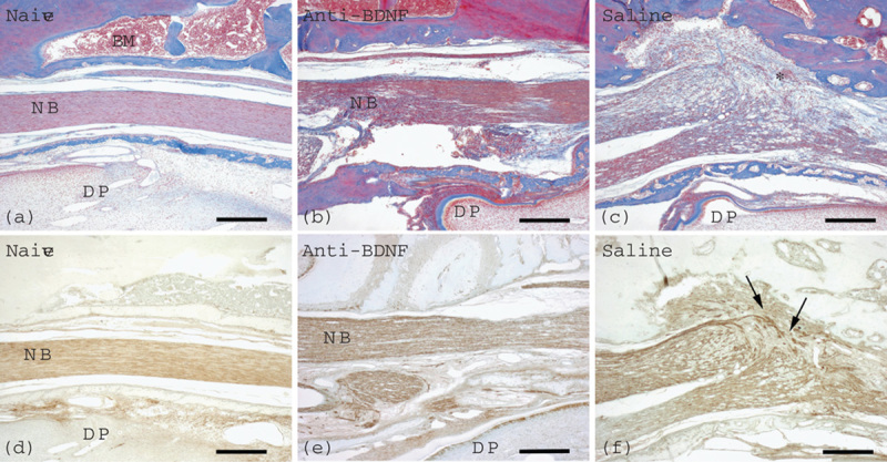
Fig. 1.

Effects of local application of an anti-BDNF antibody or physiological saline immediately after IAN transection on neuroma formation. Azan staining (a–c) and immunohistochemistry for PGP 9.5 (d–f) are presented. Samples were obtained at 2 weeks after injection of an anti-BDNF antibody (b, e) or physiological saline (c, f). In the naive group (a, d), the IAN bundle shows no damage and nerve fiber integrity as confirmed by Azan staining (a) and PGP 9.5 immunostaining (d). Neither neuroma formation nor proliferation of connective tissue is recognizable in the anti-BDNF-treated group (b, e), whereas neuroma formation with connective tissue proliferation (asterisk) is found in the vehicle control group (c, f). PGP 9.5 immunostaining shows disorganization of nerve fibers (arrows) in the vehicle control group (f), in contrast to the nerve fiber integrity in the anti-BDNF-treated group (e). Naive, anti-BDNF, and saline indicate the groups with no nerve transection, anti-BDNF antibody treatment, and nerve transection with vehicle control treatment, respectively. BDNF, brain-derived neurotrophic factor; BM, bone marrow; DP, dental pulp; IAN, inferior alveolar nerve; NB, nerve bundle; PGP 9.5, protein gene product 9.5. Scale bars=200 μm. The right and left sides in each picture indicate the proximal and the distal directions of the IAN, respectively.
