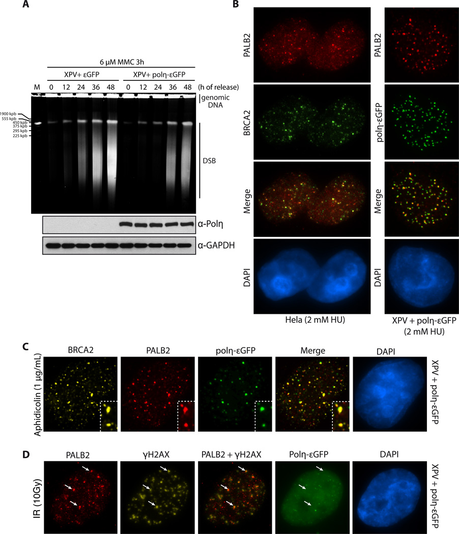
Figure 1. Polη, PALB2 and BRCA2 recruitment at replication dependent DNA double-strand breaks.
(A) Pulsed-field gel electrophoresis was used to visualize double-strand break (DSB) formation in XPV cells complemented with εGFP or Polη-εGFP, after treatment with 6 µM MMC for 3h and release for the times indicated. (B) Co-localization of PALB2 and BRCA2 or PALB2 and Polη-εGFP at DSBs induced by HU. DNA was counterstained with DAPI. (C–D) Immunofluorescence staining of the indicated DNA repair proteins at DSBs induced by aphidicolin or IR treatment in XPV cells complemented with Polη-εGFP. DNA was counterstained with DAPI. The white arrows indicate co-localization of PALB2 and γ-H2AX.

