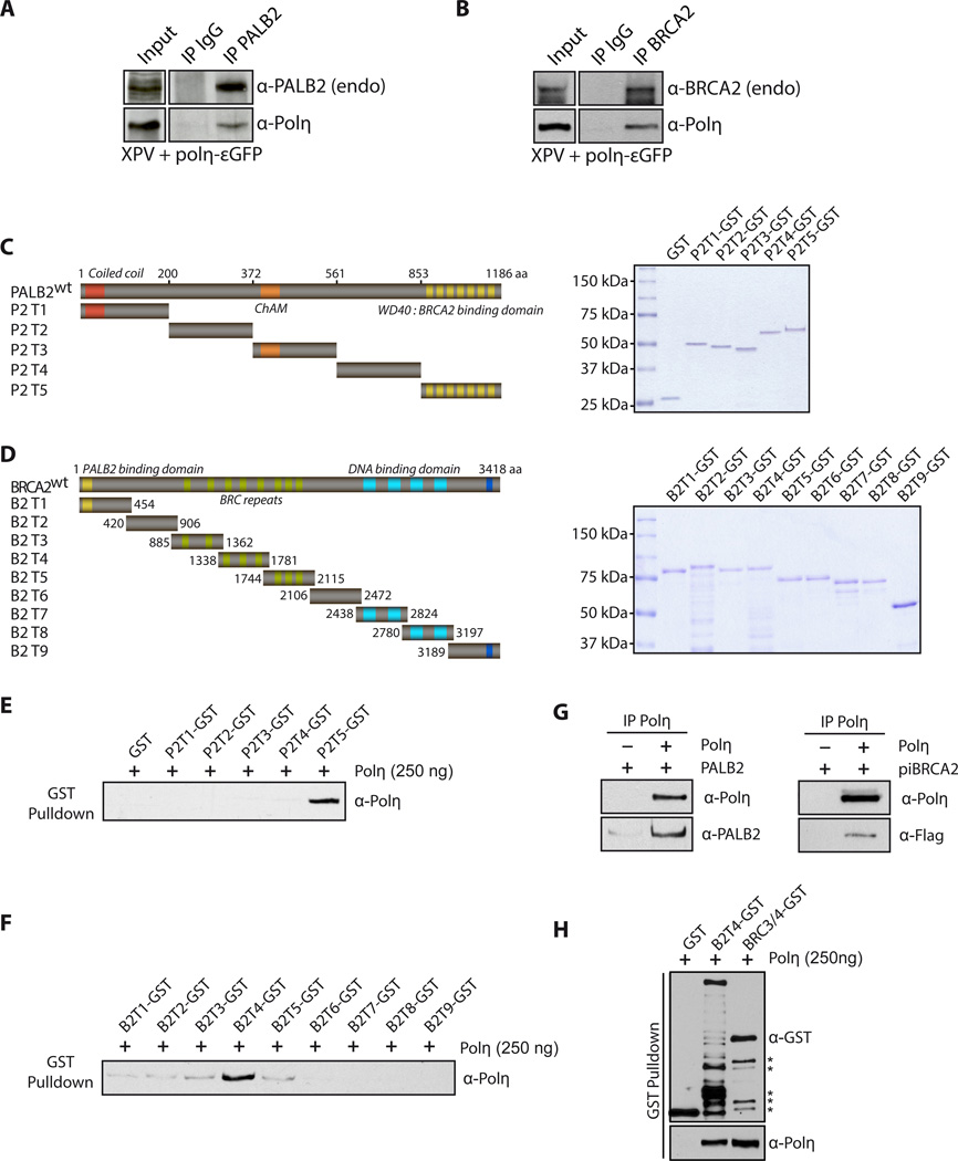
Figure 5. PALB2 and BRCA2 interact directly with Polη.
(A) Cell extracts from complemented XPV cells were subjected to immunoprecipitation with anti-PALB2 or (B) anti-BRCA2 antibodies. Immunoprecipitated proteins were detected by Western Blotting with the indicated antibodies.
(C) Left: Scheme of the PALB2 or (D) BRCA2 deletion variants fused to GST. Right: SDS-PAGE of the corresponding purified proteins. (E) GST alone or GST-PALB2 truncation (P2T1 to P2T5) were incubated with Polη followed by GST pulldown and detection of Polη by Western blotting. (F) GST alone or GST-BRCA2 truncations (B2T1 to B2T9) were incubated with Polη, followed by GST pulldown. The beads were washed and bound proteins were eluted with Laemmli buffer, and revealed by western blotting with the antibodies indicated. The input for PALB2 or BRCA2 truncations are shown in (C) and (D). (G-H) Co-immunoprecipitation of purified PALB2, piBRCA2, B2T4 or BRC3/4 and Polη. Asterisk: degradation products.

