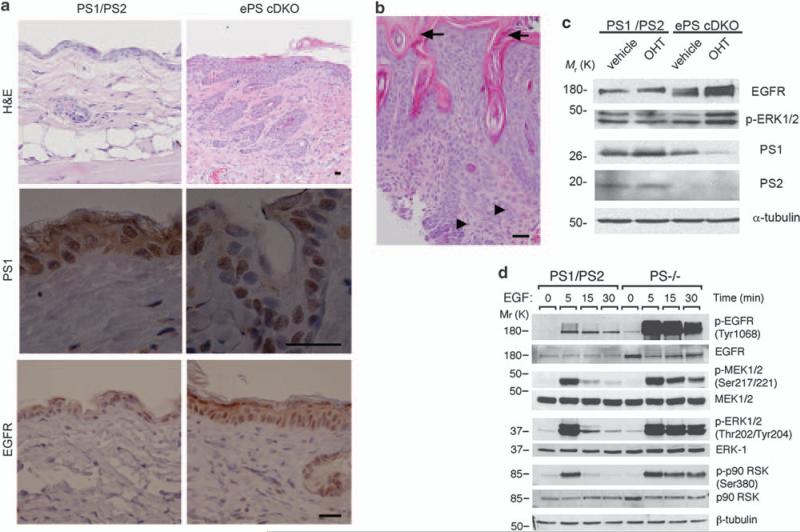
Figure 2.
PS deficiency induces epidermal hyperplasia and increased EGFR signaling in skin. (a) Paraffin-embedded sections of dorsal skin of 2.5-month-old control (PS1/PS2: PS1f/f; PS2+/–) and ePS cDKO (PS1f/f; PS2–/– ; K14-Cre-ERT2) mice were stained with hematoxylin and eosin (H&E) or processed for immunohistochemistry. H&E staining shows epidermal hyperplasia, abnormal keratinocyte proliferation and disorganization of the epidermal layer in back skin of ePS cDKO mice (× 10). Staining for PS1 (× 100) and EGFR (× 40) reveal absence of cytoplasmic and membrane PS1 expression accompanied by increased EGFR staining in epidermal keratinocytes of ePS cDKO mice. Scale bar: 30 μm. (b) H&E staining of neck skin from a 3-month-old ePS cDKO mouse showing hyperkeratosis (arrows), disorganized keratinocyte hyperplasia with infiltrating squamous epithelial cells and mitotic cells (arrowheads) consistent with histological features of squamous cell carcinomas. Scale bar: 30 μm. (c) Immunoblots showing reduced PS1 and PS2 levels and increased EGFR and phosphorylated ERK1/2 (Thr202/Tyr204) in 4-hydroxy-tamoxifen (OHT; 200 nm)-treated differentiated primary keratinocytes from ePS cDKO (PS1f/f; PS2–/– K14-Cre-ERT2) mice compared with vehicle or 4-hidroxy-tamoxifen-treated controls (PS1f/f; PS2+/–). (d) Western blotting showing increased EGFR levels and sustained EGF-induced phosphorylation of EGFR, MEK, ERK1/2 and p90RSK in PS–/– fibroblasts. The images are representative of at least three experiments.

