Figure 2.
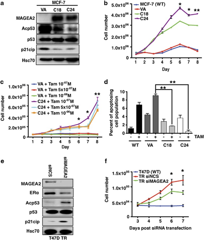
MAGEA2 overexpression is functionally linked to TR. (a) Western blot analysis of lysates (20 μg) from MCF-7-stable clones (C18 and C24) expressing exogenous MAGEA2, plus the VA control line, was probed for MAGEA2, p53, acetylated p53 and p21cip, as indicated. Blots were reprobed for Hsc70 (loading control). (b) MCF-7 (WT and VA) and clones C18 and C24 were split into six-well plates (5 × 10⁵ cells per well) and treated with 10−7M tamoxifen (TAM) 24 h later for 8 days. Triplicate wells were harvested and counted daily. (c) MCF-7 VA and C24 lines were split into 12-well plates (5 × 10⁴ cells per well) and treated with a range of tamoxifen concentrations, as indicated, over 8 days. For statistical analysis, data from all the C24 samples were averaged and compared with the averaged VA data. (d) MCF-7 control (WT and VA) and clones C18 and C24 were plated in triplicate on six-well plates (105 cells per well), grown with or without 10−7M tamoxifen for 144 h, and then assayed for Annexin V binding and PI uptake. The graph shows the percentage of early apoptotic (AnnV+PI−) cells for each line and the condition (see Supplementary Figure S4A for full analysis). (a–d) Error bars indicate the s.e. Student's t-test was used to compare data from the indicated sample with VA control; *P<0.05, **P<0.01. (e) Western blot analysis of lysates (20 μg) from T47DTR cells transiently transfected with siRNA targeting MAGEA2 or non-silencing control (NSC) for 48 h probed for the proteins indicated. (f) T47DTR cells were transfected with NSC or siRNA targeting MAGEA2. After 48 h, transfected cells and WT T47D were split into 24-well plates (3 × 10⁴ cells per well in triplicate) and treated with 10−6M tamoxifen 24 h later for 5 days. Error bars indicate the s.e. Student's t-test was used to compare data from NSC- and siMAGEA2-transfected cells; *P<0.05.

