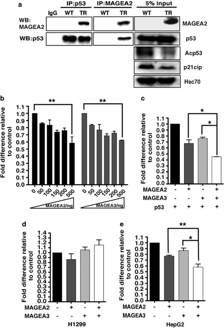
Figure 3.
MAGEA2 interacts with the p53 pathway in breast cancer lines. (a) Whole-cell extract (WCE) lysates from WT and TR T47D cells were immunoprecipitated (IP) for either p53 (left panels) or MAGEA2 (middle panels), and subsequent western blots (WBs) were probed for the same proteins, as indicated. Separate WBs (loaded with 5% of the protein levels used for the IP experiments; right panels) were probed for the indicated proteins. (b) H1299 (p53-null) cells were transfected with the pG1PyLuc reporter plasmid (150 ng), a p53 expression vector (10 ng) and increasing amounts of MAGEA2 or MAGEA3 expression vectors, as indicated. (c) As in (b) but with MAGEA2 and/or MAGEA3 expression vectors (100 ng), as indicated. (d and e). As in (c) but with the p21cip-Luc reporter plasmid (150 ng) without the addition of p53 expression plasmid using p53-null H1299 cells (d) or p53 WT HepG2 cells (e). (b–e) Cells were harvested after 48 h. Luciferase values were corrected for transfection efficiency and expressed as fold activity compared with the reporter alone control (set at 1) using data from three independent experiments, each performed in triplicate; error bars indicated the s.e. Student's t-test was used to compare data from the indicated samples, *P<0.05; **P<0.01.

