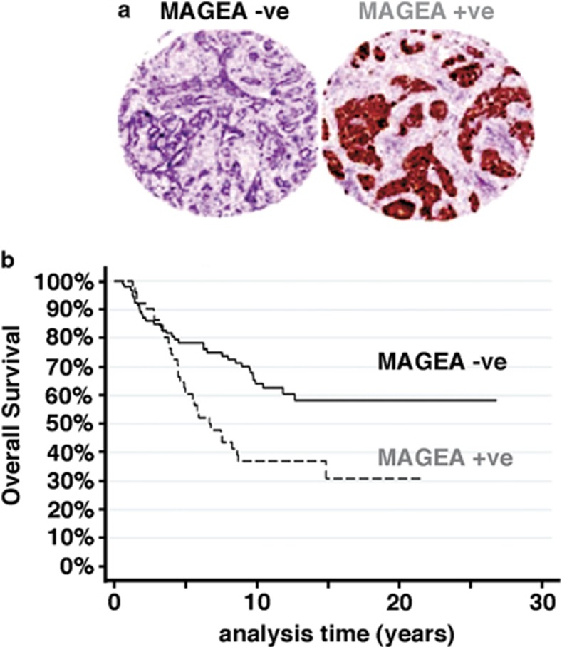
Figure 6.
MAGEA expression in ER+ tumors correlates with reduced OS. Expression of pan-MAGEA antigens was assessed in 144 cases of ER+, tamoxifen-treated primary breast cancer by immunohistochemical analysis on whole sections or TMA, as available. (a) Representative patterns of positive and negative staining on TMA (x5 magnification). (b) Kaplan–Meier survival curves showing the relationship between positive and negative staining for MAGEA on OS. The two-sided P-value (P=0.006) was calculated using log-rank testing.

