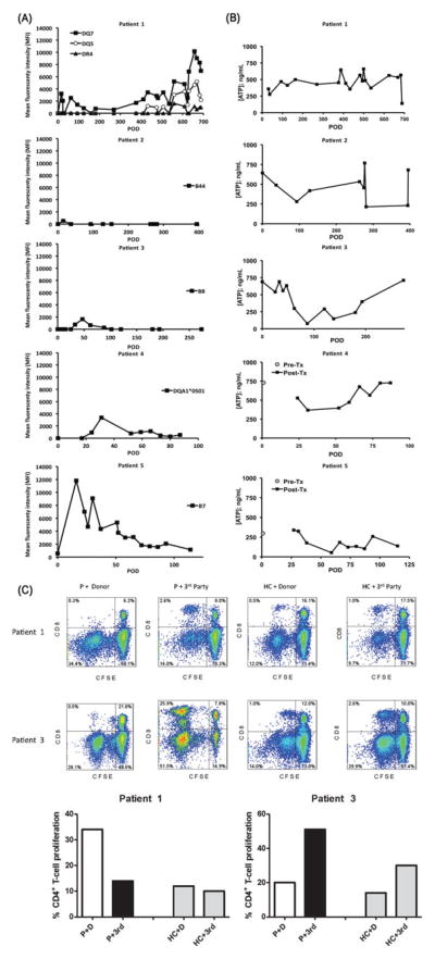
FIGURE 3.
A, Both human leukocyte antigen class I and class II donor-specific alloantibodies were first observed within the first month after transplant. Changes in donor-specific alloantibodies were limited to specific haplotypes in each patient, except for patient 1, who initially showed donor-specific alloantibodies against DQ7, which resolved with treatment but later recurred together with DR4 and DQ5 donor-specific alloantibodies. Such changes coincided with admitted noncompliance with immunosuppression. The donor-specific alloantibodies significantly subsided after compliance was re-established. B, Global CD4+ T-cell immune response were measured using the Immuknow™ assay and categorized into low (<225 ng/mL ATP), moderate (226–525 ng/mL ATP), and high (>525 ng/mL ATP). Patients 1, 2, and 4 demonstrated moderate responses over a majority of time points. In patients 2 and 3, the assay indicated a stronger cellular immune reactivity early after transplantation, with a dip during 12–18 months (patient 2) and 3 to 6 months (patient 3). However, recent values have trended to pretransplant levels. In patient 5, the response has varied between low and moderate despite stable target troughs on low-dose tacrolimus monotherapy. C, One-way carboxyfluorescein diacetate succinimidyl ester-MLR to assess T-cell allospecific proliferation. Proliferation of alloreactive CD3+ T cells was measured by carboxyfluorescein diacetate succinimidyl ester dilution (% of carboxyfluorescein diacetate succinimidyl ester -low cells) of CD4+ and CD8+ T cells after 5 days of in vitro stimulation of patient or healthy control peripheral blood mononuclear cells with donor (D) or third (3rd)-party peripheral blood mononuclear at 1:1 ratio. FACS analysis revealed that patient 1 displayed a significant CD4+ T-cell proliferation to donor antigens as opposed to third-party stimulation (donor-specific reactivity). Patient 3 demonstrated CD4+ and CD8+ T cells with normal memory subset distribution, a robust CD4+ and CD8+ T-cell proliferation to third-party stimulation, but lower levels of CD4+ and CD8+ T-cell proliferation to donor stimulation, denoting a quiescent status (donor-specific hyporesponsiveness).

