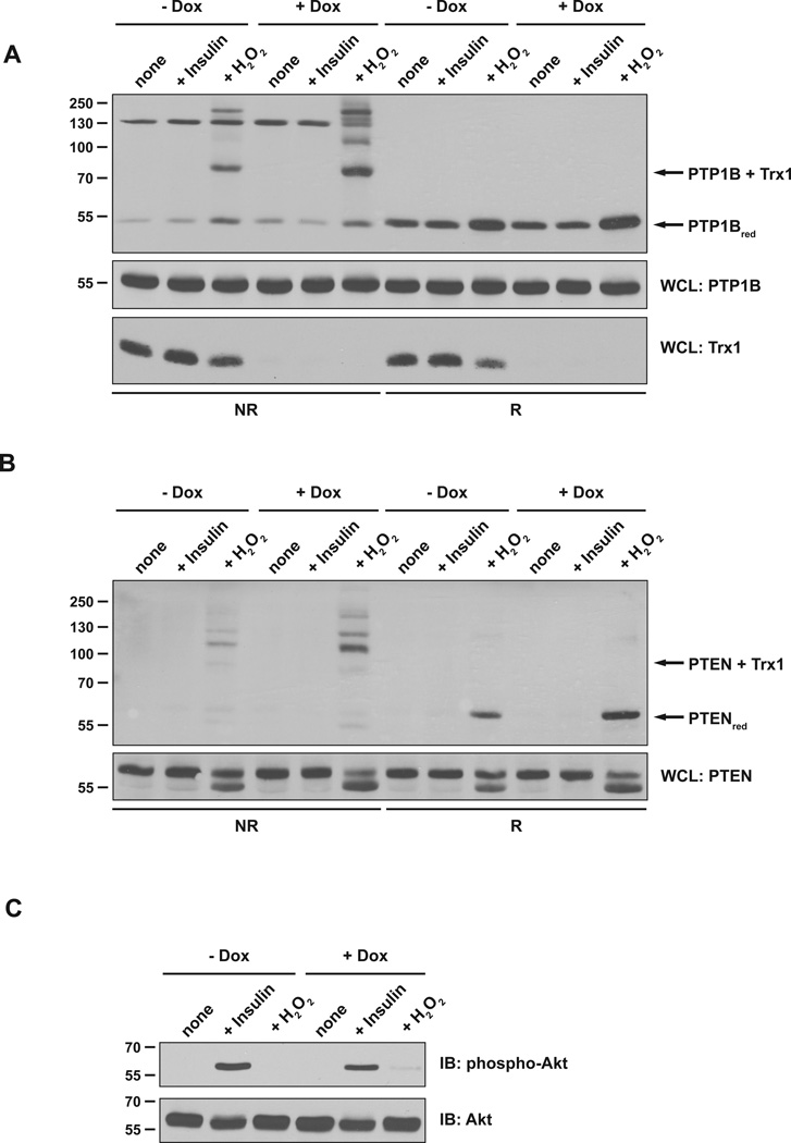
Figure 4. TRX1 contributed to reduction of PTEN and PTP1B in cells.
(A) HeLa T-REx-252 cells were either left untreated or treated with 1 µg/ml doxycycline (Dox) for 5 days. Cells were stimulated with 100 nM insulin or 1 mM H2O2 and lysate samples were incubated with the TRX1 trapping mutant (TRX1-C35S). TRX1-substrate complexes (upper panel) and samples of WCL (middle panel) were resolved by non-reducing and reducing SDS-PAGE and analyzed by anti-PTP1B immunoblotting. The disulfide-linked TRX1-PTP1B complex and reduced PTP1B are indicated. WCL samples were analyzed by TRX1-specific immunoblotting to verify TRX1 depletion (lower panel).
(B) Samples as in (A) were analyzed by PTEN-specific immunoblotting. The disulfide-linked TRX1-PTEN complex and reduced PTEN are indicated. The markers indicate kDa.
(C) Samples of WCL were analyzed with anti-phospho-Akt (Ser473) antibody to confirm responsiveness of the cells to insulin.

