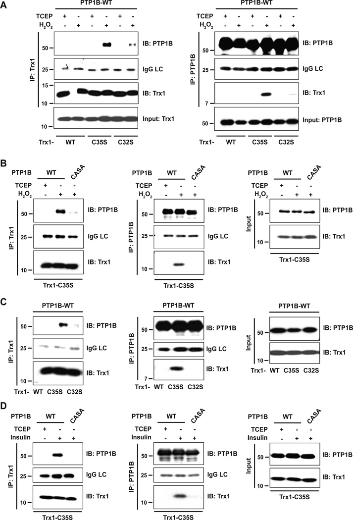
Figure 5. An ectopically expressed TRX1 trapping mutant co-precipitated with PTP1B after insulin stimulation.
(A) Different His6-tagged TRX1 constructs were co-expressed with Flag-tagged PTP1B in 293T cells. Cells were treated with 1 mM H2O2 to oxidize intracellular proteins. Untreated cells were harvested in the presence of TCEP. TRX1 (left panel) or PTP1B (right panel) was immunoprecipitated from whole cell lysates and analyzed by immunoblotting. TRX1 and PTP1B expression was confirmed in input lysates. LC: IgG light chain
(B) TRX1-C35S and PTP1B-WT or the CASA mutant were co-expressed in 293T cells. TRX1 (left panel) or PTP1B (middle panel) was immunoprecipitated from lysates of reduced (TCEP) or H2O2-treated cells and immunoprecipitates were analyzed as described in (A). TRX1 and PTP1B expression was confirmed in input lysates (right panel).
(C) 293T cells co-expressing different His6-tagged TRX1 constructs and Flag-tagged PTP1B were treated with 100 nM insulin for 10 minutes, the time point at which insulin receptor β subunit (IRβ) and IRS-1 displayed maximum tyrosine phosphorylation [16]. TRX1 (left panel) or PTP1B (middle panel) was immunoprecipitated from whole cell lysates and immunoprecipitates were analyzed as described in (A). TRX1 and PTP1B expression was confirmed in input lysates (right panel).
(D) 293T cells co-expressing TRX1-C35S and PTP1B-WT or -CASA were treated with 100 nM insulin. PTP1B-WT or -CASA were immunoprecipitated and analyzed as described in (A). TRX1 and PTP1B expression was confirmed in input lysates (right panel).

