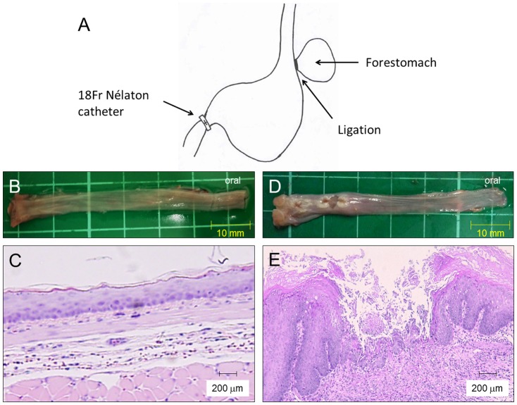
Figure 1. Induction of rat acid reflux esophagitis, macroscopic, and histological findings.
(A) A scheme of a rat reflux esophagitis model. (B) Macroscopic appearance of a normal esophagus. (C) Histology of the normal esophagus revealed thin epithelium with few inflammatory cells. (D) Macroscopic appearance of reflux esophagitis showed several erosions and ulcers at the middle and lower esophagus. (E) Mucosal thickening with basal cell hyperplasia and marked inflammatory cell infiltration were observed in reflux esophagitis. (Hematoxylin-eosin staining, original magnification ×200).

