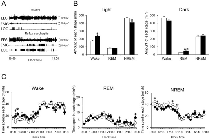
Figure 3. Effect of reflux esophagitis on sleep.
(A) Typical EEG, EMG, and LOC of control and reflux esophagitis. (B) Effect of reflux esophagitis on the amount of each stage during the 12-h light period and 12-h dark period. (C) Time course analysis of each stage during a whole day. N = 8. Data are mean ± SEM. White bars and circles represented control rats. Black bars and circles represented reflux esophagitis rats. *p<0.05 versus control. **p<0.01 versus control. EEG, electroencephalograph; EMG, electromyography; LOC, locomotor; NREM, nonrapid eye movement; REM, rapid eye movement.

