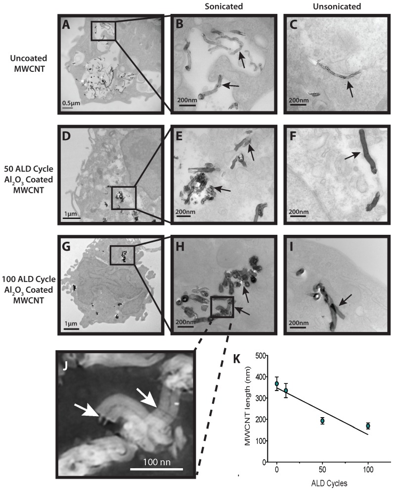
Figure 2. Transmission electron microscopy of human macrophages engulfing MWCNTs.
A) Lower magnification (x18000) of uncoated, sonicated MWCNTs contained in vesicles within a THP-1 macrophage. B) Higher magnification (x56000) of uncoated, sonicated MWCNTs within a vesicle of a THP-1 macrophage. C) High magnification (x56000) of a single (unsonicated) MWCNT within the cytoplasm of a THP-1 macrophage. D) Lower magnification (x7100) of a THP-1 macrophage with sonicated Al2O3 coated MWCNTs (50 ALD cycles) in the cytoplasm. E) Higher magnification (x56000) of a cluster of sonicated Al2O3-coated MWCNT fragments (50 ALD cycles within the cytoplasm of a THP-1 macrophage. F) High magnification (x36000) of a THP-1 macrophage with Al2O3 coated MWCNTs (50 ALD cycles –unsonicated) in the cytoplasm in close proximity to the nucleus. G) Low magnification (x7100) of a macrophage with Al2O3-coated (100ALD cycles) sonicated MWCNTs in the cytoplasm. H) Inset showing a higher magnification (x56000) of a cluster of sonicated Al2O3-coated MWCNTs (100 ALD cycles) within the cytoplasm of a macrophage. I) High magnification (x56000) showing unsonicated Al2O3-coated (100 ALD cycles) MWCNTs in the cytoplasm of a THP-1 macrophage. J) High magnification inverted image of ALD-coated MWCNTs from panel H showing fracture points of breakage (arrows). K) Al2O3 coated MWCNT length (nm) as a function of ALD cycle post sonication. Data represent mean values (±SEM) of 20 measurements of nanotubes thickness and length.

