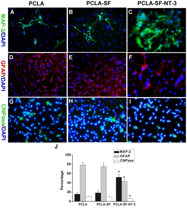
Figure 4. Differentiation of NSCs in vitro.
(A–I) Cells cultured on PCLA (A, D, G), PCLA-SF (B, E, H), and PCLA-SF-NT-3 (C, F, I) membrane were immunostained with MAP-2 for neurons (A–C), GFPA for astrocytes (D–F), CNPase for oligodendrocytes (G–I). The nuclei were stained with DAPI. (J) Quantitative analysis of the NSC differentiation in vitro. Scale bars = 25 µm, *P<0.05; n = 4.

