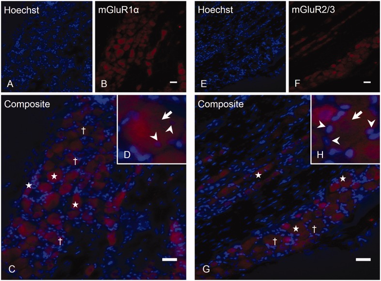
Figure 2.
Expression of mGluR1α and mGluR2/3 in the TG. Labeling TG sections with rabbit anti-mGluR1α (B) and rabbit anti-mGluR2/3 (F), followed by counterstaining with Hoechst (A, E), revealed both positive (stars) and negative (cross) neurons (exemplified in 2C and G). Higher magnification of two positive neurons are shown for each mGluR staining (D, H, mGluR1α and mGluR2/3, respectively), where SGCs (arrowheads) were located inside the immunoreactive area (arrow) of the mGluR1α staining (D), indicating that trigeminal SGCs express mGluR1α. In contrast, SGCs (arrowheads) were located outside the immunoreactive area (arrow) of the mGluR2/3 staining (H), indicating that these SGCs do not express mGluR2/3. Magnification = 200× (A, B, C, E, F and G). Scale bars = 50 µm. mGluR, Metabotropic glutamate receptor; TG, trigeminal ganglion; SGCs, satellite glial cells.

