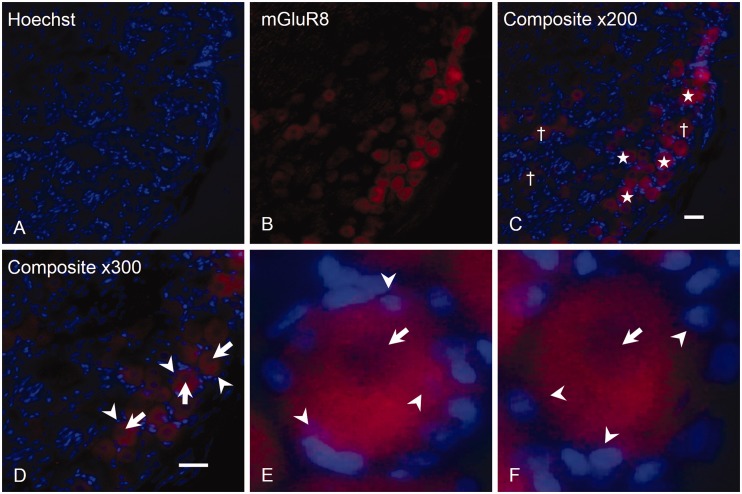
Figure 3.
Expression of mGluR8 on trigeminal neurons and SGCs. TG sections were immunostained with rabbit anti-mGluR8 (B) followed by nuclear stain Hoechst (A). A merge (C) is shown, exemplifying positive (stars) and negative (cross) neurons. The same section at 300× magnification (D) shows the characteristic surrounding of neurons (arrows) by SGCs (arrowheads). (E) A positive neuron at higher magnification, where SGCs (arrowheads) are located within the immunoreactive area (arrow), indicating that these SGCs express mGluR8, whereas (F) shows another positive neuron, with SGCs (arrowheads) located outside the immunoreactive area, indicating that these SGCs do not express mGluR8. Magnification = 200× (A, B and C) and 300× (D). Scale bars = 50 µm. TG, Trigeminal ganglion; mGluR: metabotropic glutamate receptor; SGCs: satellite glial cells.

