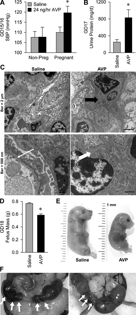
Figure 3. Infusion of vasopressin during pregnancy is sufficient to cause the cardinal phenotypes of preeclampsia in mice.
(A) Systolic blood pressure on gestational days 15 and 16. Non-pregnant mice infused with saline (n=15) or AVP at 24 ng/hr (n=5) for 15–16 days, and pregnant mice infused with saline (n=16) or AVP (n=11). * P<0.05 versus all other groups. (B) Total protein lost to urine per day, on gestational day 17. Pregnant mice infused with saline (n=5) or AVP at 24 ng/hr (n=9). * P<0.05 versus saline. (C) Electron micrographs of renal cortex, illustrating glomerular endotheliosis. Left panels are from a saline infused animal which had a glomerular basement membrane thickness within normal limits (thin white arrow). Right panels are from animals that received vasopressin infusion (24 ng/hr). Redundant endothelial cell membrane is present (thick black arrow), and basement membranes are moderately to markedly thickened with electron dense material (thick white arrow). (D) Average GD18 fetus mass from mice infused with saline (117 fetuses from 16 pregnancies) or AVP at 24 ng/hr (115 fetuses from 16 pregnancies). * P<0.05 versus saline. (E) Photographs of representative gestational day 18 fetuses from pregnant dams infused with saline and 24 ng/hr AVP, illustrating intrauterine growth restriction. (F) Photographs of feto-placental units in utero on gestational day 18 during 24 ng/hr AVP infusion. White arrows indicate resorbed feto-placental units.

