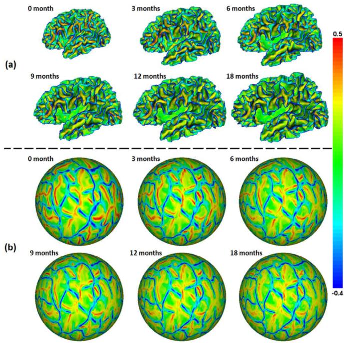
Fig. 1.
(a) Longitudinal inner cortical surfaces of the left hemisphere of a representative infant from 0 to 18 months of age, color-coded by the mean curvatures. (b) Group-wisely aligned longitudinal spherical surfaces of the left hemisphere of the infant, color-coded again by the mean curvatures. Red colors indicate sulci, and blue colors indicate gyri.

