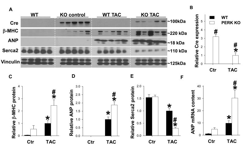
Figure 3. PERK KO exacerbated TAC-induced changes in myocardial Serca2a, ANP and β-myosin heavy chain (β-MHC) expression.
Tissue was collected from WT and PERK KO mice 5 weeks after TAC or control conditions, and lysates were examined by western blot for expression of Cre (A, B), atrial natrurietic peptide (ANP) (A, C), β-MHC (A, D) and Serca2a (A, E). Vinculin was used as a loading control. Relative ANP mRNA content in each group (F). * indicates p<0.05 comparing TAC to control. # indicates p<0.05 comparing WT to PERK KO.

