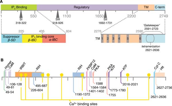
Figure 2. Topology of structure-functional domains in primary sequence of IP3R.
(A) Four key regions defined in the primary structure of IP3R are highlighted: the N-terminal ligand-binding region, the large central modulatory region, the membrane-spanning/pore-forming, and the C-terminal ‘gate-keeping’ region; SI, SII and SIII refer to the three splice sites. Lower panel shows detailed topology of domains within the N- and C-terminal regions (see also Figure 5). (B) Putative binding sites for several modulatory molecules of IP3R are indicated in the primary sequence of the receptor protein: CaM – calmodulin, IRBIT- IP3R binding protein released with IP3, FKBP12 - 12-kDa FK506-binding protein, PKA – cAMP-dependent protein kinase A, Cyt c – cytochrome c; Htt – huntingtin; RIH - RyR/IP3R homology regions. Amino acid residue numbering is the same as for the mouse IP3R1 (GI: 313104120) [1].

