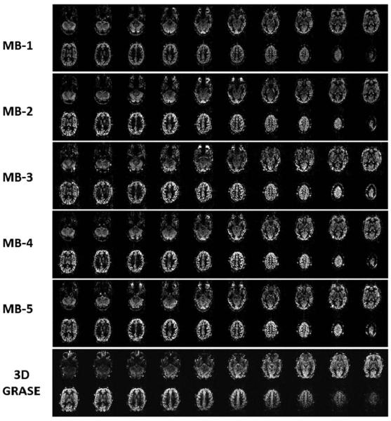
FIG. 4.
Comparison of perfusion weighted images covering 120-mm slice volume, whole-brain coverage, in the second subject. The 20 slice coverage with MB-1 (standard EPI), MB-2, MB-3, MB-4, and MB-5 slice acceleration factors. The 3D GRASE ASL images are shown offset by one image to the right of the corresponding positioned SMS-EPI ASL images.

