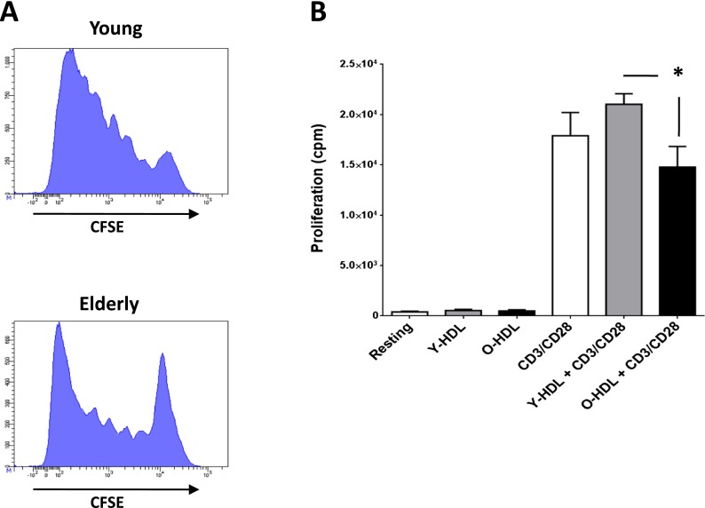
Fig. 5.
Impact of aging and HDL on T cell proliferation. a As aging has been associated with loss of functionalities, we tested the proliferative capacity of CD3+ T cells from young and elderly individuals and show the CFSE dilution assay of a typical experiment. PBMCs were stimulated with αCD3αCD28 antibodies for 5 days and stained for CD3 before FACS analysis. T cells from young individuals show a high proliferative response that is lower in the case of elderly individuals. b The proliferative response of T cells from elderly individuals following αCD3αCD28 stimulation was also tested in the presence of HDL as a putative immunomodulatory agent. Proliferation was tested by addition on 3H-thymide for the last 4 h of the proliferation assay and represented as counts per minute (cpm). The only significant difference observed (*p < 0.05) is the difference between T cells treated with HDL from young versus old individuals

