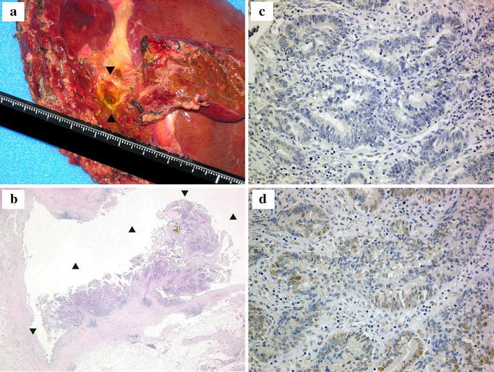
Fig. 3.

The results of a histopathological analysis of the tumor sample obtained from the left lobectomy. a The gross appearance of the resected tissue. b The H&E staining (×100) revealed a well-differentiated papillary hilar bile duct cancer [T1 (fibromuscular layer, fm) N0M0 Stage IA (Union Internationale Contre le Cancer, UICC TNM staging) (arrowheads)]. c Immunostaining of the tumor for MLH1 (×400). d Immunostaining of the tumor for MSH2 (×400). Immunoreactivity was observed for MSH2, but not for MLH1
