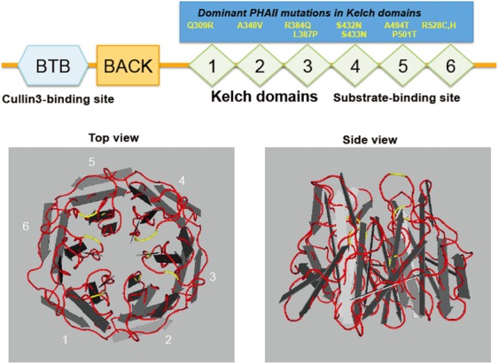
Figure 3. Structure of Kelch-like proteins.

The upper panel shows the structure of Kelch-like (KLHL) proteins with N-terminal BTB and BACK domains and five to six C-terminal Kelch domains, and most autosomal dominant mutations causing pseudohypoaldosteronism type II (PHAII). The BTB domain is a binding site for Cullin 3 and Kelch repeats constitute a propeller structure, as shown in the lower panels, and capture a substrate. Each Kelch domain forms a blade, and most PHAII-causing mutations (shown in yellow lines) are located in the loop regions linking each blade, which may be involved in substrate binding.
