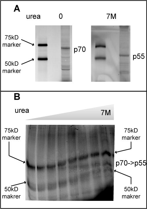
Figure 4. Conversion of p70 into p55 in the presence of urea.

(A) CLICK reacted lysates from H2122 cells treated with P7C3-S326 were separated on a standard 8% SDS-PAGE gel (left panel) or one supplemented with 7M urea (right panel), and visualized on a Typhoon scanner. Asterisk indicates the crosslinked protein that migrates as 70KDa in a standard SDS-PAGE gel and 55KDa in the SDS-PAGE gel supplemented with 7M urea. (B) The same lysates were resolved on a horizontal urea gradient gel co-loaded with crosslinked p70 and the same 75kD and 50kD size standard proteins displayed in panel A. The horizontal urea gradient proceeds from zero denaturant on the left to 7M denaturant on the right.
