
Figure 2. Poly-ubiquitination by Hrd1p.
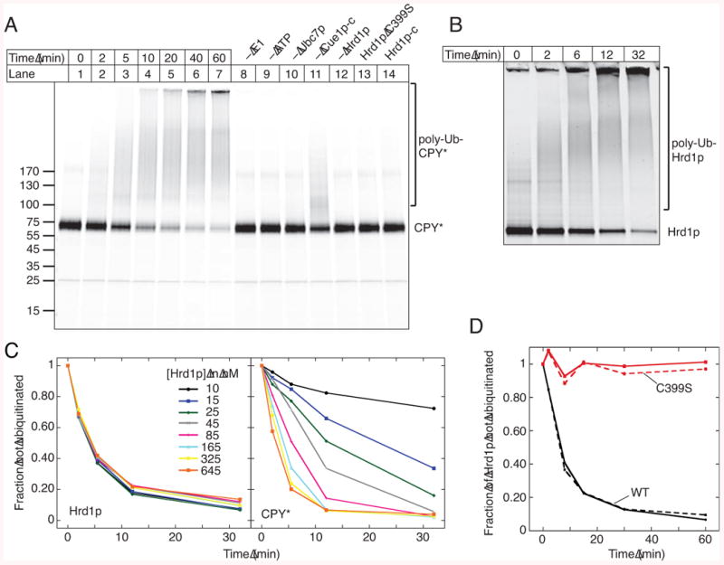
(A) Time-course of ubiquitination of CPY* labeled with DyLight800. Some reactions were analyzed after 60 min with the indicated components omitted. Where indicated, wild type Hrd1p (100 nM) was replaced with 100 nM of an inactive Hrd1 p mutant (C399S) or 1 μM of the cytoplasmic domain of Hrd1 p (Hrd1p-c).
(B) Time-course of auto-ubiquitination of Hrd1p labeled with DyLight680.
(C) The time-course of auto- and substrate-ubiquitination was determined in parallel. The concentration of labeled Hrd1p was kept constant, while that of unlabeled Hrd1p was varied.
(D) The time-course of auto-ubiquitination was determined with 50 nM of labeled wild type (WT) Hrd1p or C399S mutant in the absence or presence of a 10-fold excess of unlabeled WT Hrd1p (solid and broken curves, respectively).
See also Figure S2.