
Fig. 1.
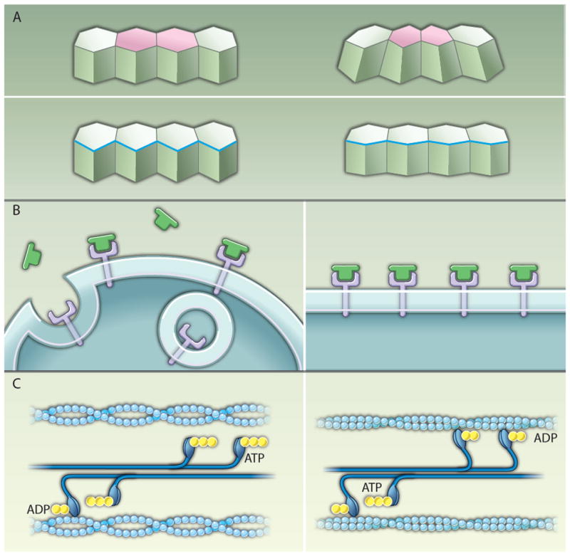
Mechanisms of tension-mediated actomyosin regulation. (A) Actomyosin contraction promotes changes in cell shape during epithelial morphogenesis. During mesoderm invagination and in the amnioserosa during dorsal closure, contraction of an apical actomyosin network (pink) drives apical constriction (top). Leading-edge cells of the lateral epidermis align through the contraction of a multicellular cable (blue) that contributes to dorsal closure (bottom). (B) A model of the plasma membrane as a mechanosensor. In the absence of tension, Fog (green) signaling through its putative receptor (purple) is proposed to be counteracted by endocytosis (left). When the plasma membrane is subjected to external tension, the bending and pinching off of vesicles is inhibited, promoting Fog signaling (9) (right). (C) A model of the myosin II motor as a mechanosensor. For simplicity only one head of each myosin motor is shown. When actin filaments (light blue) are under low tension, myosin II motors in bipolar filaments (dark blue) are rapidly released after adenosine triphosphate (ATP) (yellow) hydrolysis and the power stroke (left). In actin filaments under tension, the ADP bound form of myosin II is stabilized (34-36), leading to the accumulation of myosin II molecules on the actin filament (right).