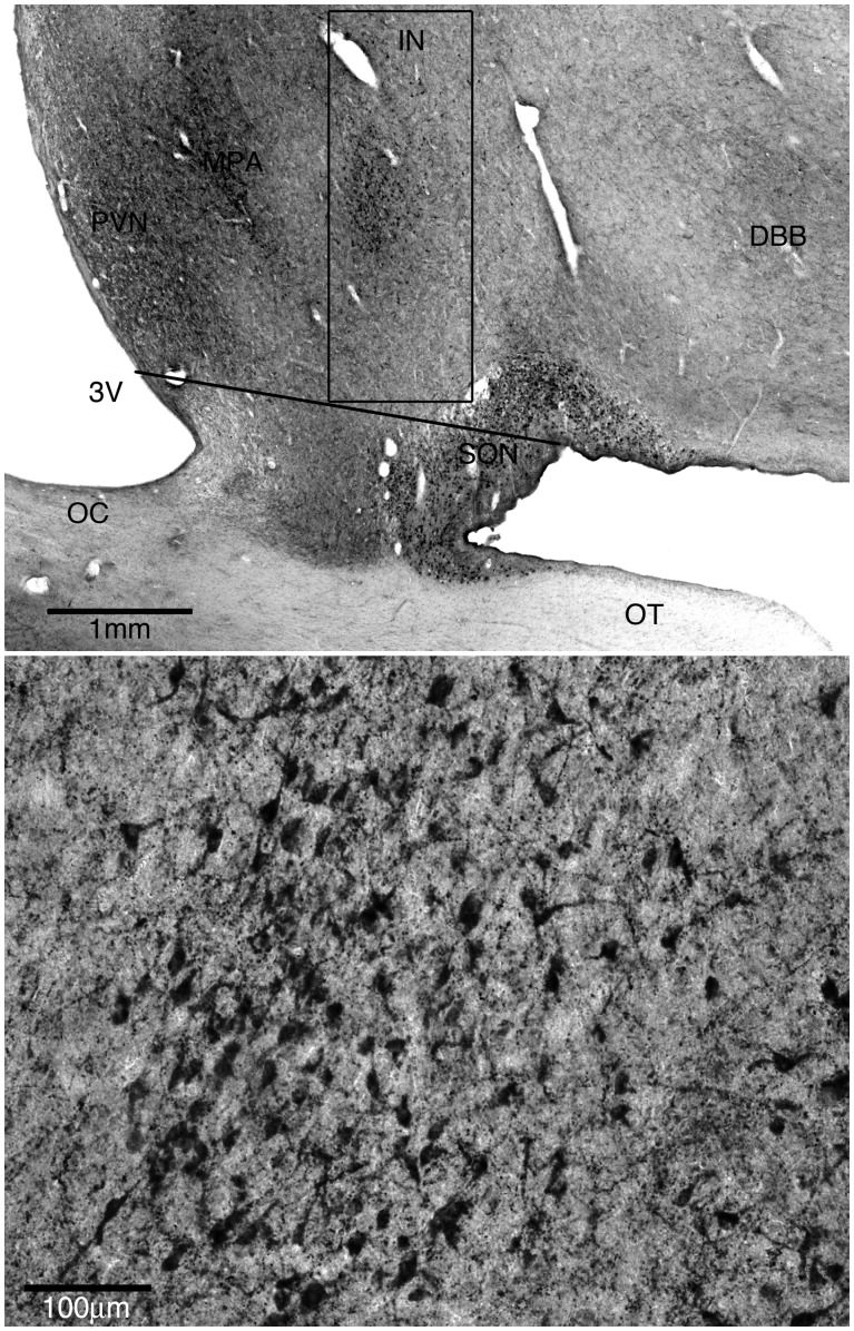
Figure 1.
The intermediate nucleus of the hypothalamus. (A) Representative low power view of the anterior hypothalamus stained immunohistochemically for galanin. The box indicates the counting frame used to define the intermediate nucleus. The long axis of the box is parallel to the lateral wall of the third ventricle. The inferomedial corner of the box is at the midpoint of a line, parallel to the base of the brain, connecting the angle between the base of the brain and the chiasmatic region inferolateral to the supraoptic nucleus to the lateral wall of the third ventricle. 3V = third ventricle; IN = intermediate nucleus; DBB = nucleus of the diagonal band of Broca; OT = optic tract; OC = optic chiasm. (B) Representative high power view of intermediate nucleus of the hypothalamus stained for galanin.

