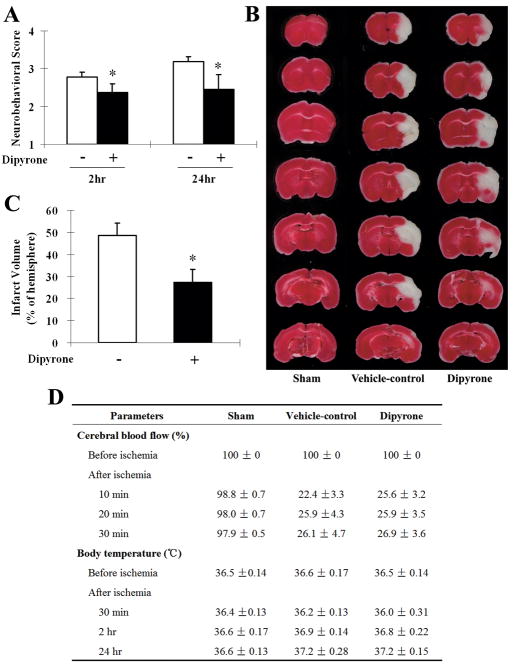
Figure 5.
Improvements in neurological behavior (A) and infarct volume (B, C) of intracerebroventricularly (i.c.v.) injection of dipyrone in the mouse model of focal cerebral ischemia. Cerebral ischemia was induced by intraluminal suture occlusion of middle cerebral artery. Dipyrone (100 ng) was i.c.v. delivered 30 min before the onset of MCAO. A, Neurobehavioral scores were assessed 2 and 24 hours after ischemia as scale described in the Methods section. B, The TTC-stained coronal brain sections are from representative animals of sham-operated group, vehicle-control group and dipyrone-treated group, respectively. Brains were removed after 24 hours of MCAO, and coronal sections thereof were stained with 2% TTC. The infarcted tissue is white, whereas live tissue is darkly stained by TTC. C, Brain infarct volume was determined by serial reconstruction from coronal sections prepared by staining with 2% TTC 24 hours after ischemia as in B. D, Physiological parameters were monitored before and after the onset of ischemia. No statistical difference was found in body (rectal) temperature between the vehicle and the dipyrone-treated group. Data are presented as mean ± S.E.M. (n=10). *, P < 0.05 versus vehicle-control group.

