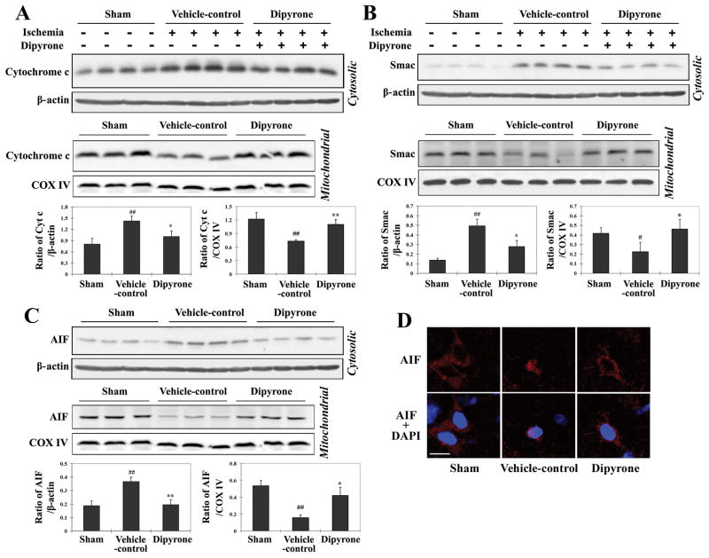
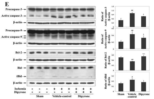
Figure 7.
Dipyrone delivered by intracerebroventricular (i.c.v.) injection inhibits molecular events associated with cell death in ischemic brain tissue. Cerebral ischemia was induced by intraluminal suture, thereby occluding the middle cerebral artery. 100 ng of dipyrone was administered by i.c.v. injection 30 minutes before the onset of MCAO. After 12 hours of ischemia, brains were removed and ischemic territories were isolated. Cytosolic fractions, mitochondrial fractions or total lysates were prepared by homogenization and centrifugation as described in the Methods section. A–C, Samples of the cytosolic (n=4) and mitochondrial (n=3) fractions were analyzed by western blot using antibodies to cytochrome c (A), Smac (B), and AIF(C). D, The release and nuclear redistribution of AIF in the brain after ischemia. Scale bar, 10 μm. E, Samples of total lysate were analyzed by western blot with antibodies to pro- and active caspase-3, caspase-9, Bcl-2, and Bid (n=3). β-actin or COX IV staining was used as an internal loading control to the cytosolic fractions/total lysates or mitochondrial fractions, respectively. Each lane in the blots represents a separate mouse. The bar graphs were generated by densitometry, and the values are presented as mean ± S.E.M. #, P < 0.05; ##, P < 0.01 versus sham-operated group. *, P < 0.05; **, P < 0.01 versus vehicle-control group.


