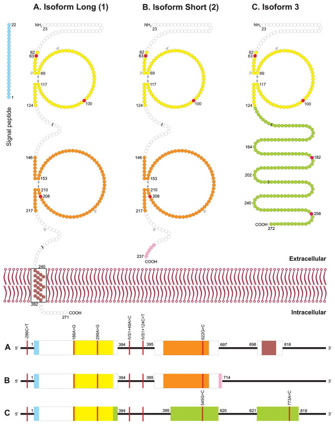
Figure 1. ICAM4 protein and ICAM4 pre-mRNA.
Schematic models of ICAM4 protein are depicted for the 3 known isoforms (upper panel). Variations were found at 3 amino acid positions (red circles). The first 22 amino acid positions are predicted to be a signal peptide (blue circles). Additional predicted structural features are the 2 immunoglobulin (Ig) domains (yellow and orange circles) and a transmembrane segment (brown circles). Isoform Long (1) is a single-pass transmembrane protein (A), while Isoform Short (2) and Isoform 3 are secreted (B and C). Some protein segments in isoforms 2 and 3 differ from isoform 1 (purple, pink and green circles). The projections on circle surfaces denote the positions of the 4 N-glycosylation sites. The 7 variant nucleotide positions are depicted in the 3 pre-mRNA ICAM4 isoforms (bottom panel). Three variants were found in the exons (boxes) and 4 in the introns (lines). The nucleotide stretches of the exons are colored according to the encoded protein segments. The exon boundaries in the ICAM4 cDNA, as reflected in the amino acid sequence, are indicated (black bars).

