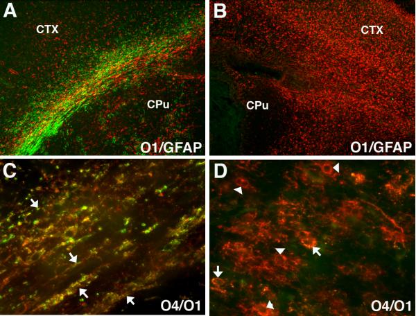
Figure 3.
Numerous late oligodendrocyte progenitors (preOLs) accumulate in chronic myelin-deficient perinatal white matter lesions. Lesions were generated in response to unilateral hypoxia-ischemia in the postnatal day 3 rat with the contralateral hemisphere serving as control (see Segovia et al., 2008). (A) Normal early myelination (O1-antibody; green) in control subcortical white matter (corpus callosum/external capsule) at P10 is seen with low levels of GFAP-labeled astrocytes (red) mostly concentrated over the white matter. (B) Absence of myelin in the contralateral post-ischemic lesion coincided with a diffuse glial scar that stained for GFAP-labeled astrocytes. (C) Early myelination in control white matter at P10 with sheaths (yellow) double-labeled for O4 and O1 antibodies. (D) Absence of myelin in the contralateral lesion coincided with clusters of preOLs (O4+O1-) in maturation arrest (red; arrowheads). Such dense clusters of preOLs are not normally seen in control white matter and are consistent with the pronounced proliferative state that is triggered in response to injury. Peak preOL density can expand roughly 4-fold relative to control. Oligodendroctes (yellow; arrows; O4+O1+) are rarely seen in the lesions. Abbreviations: CTX, cerebral cortex; CPu, caudate putamen.

