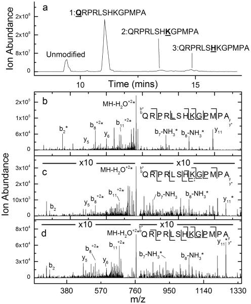
Figure 1.
(a) Total ion chromatogram after HPLC separation of DEPC-labeled apelin 13. (b) CID spectrum of the (M+2H)2+ ion of the second chromatographic peak from the DEPC-labeled sample of apelin 13, indicating that the N-terminus is modified. (c) CID spectrum of the (M+2H)2+ ion of the third chromatographic peak from the DEPC-labeled sample of apelin 13, indicating that Lys8 is modified. *Interfering ions make assignment of the labeled and unlabeled versions of the y6 ion somewhat ambiguous. (d) CID spectrum of the (M+2H)2+ ion of the fourth chromatographic peak from the DEPC-labeled sample of apelin 13, indicating that His7 is modified.

