
Figure 1.
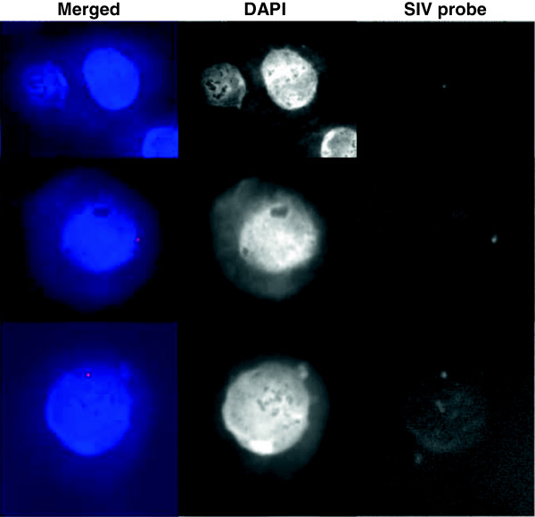
Halo-FISH analysis of acutely SIV-infected C8166 T cells. Integration of SIV into the genomic DNA of the C8166 T cell line. Three acutely SIV-infected cells were shown in three different rows. Halo-FISH analyses of cells 1 d after acute infection was performed by using a SIV gag-env DNA probe. Nuclei were visualized by DAPI staining. Single integration events of SIV proviral DNA (SIV probe, white spots; Merged with DAPI staining, red spots) into the nuclear matrix at regions of high transcriptional activity (blue sphere) are clearly visible. No integration into the associated HALO (regions of low transcriptional activity surrounding the nuclear matrix) was observed.