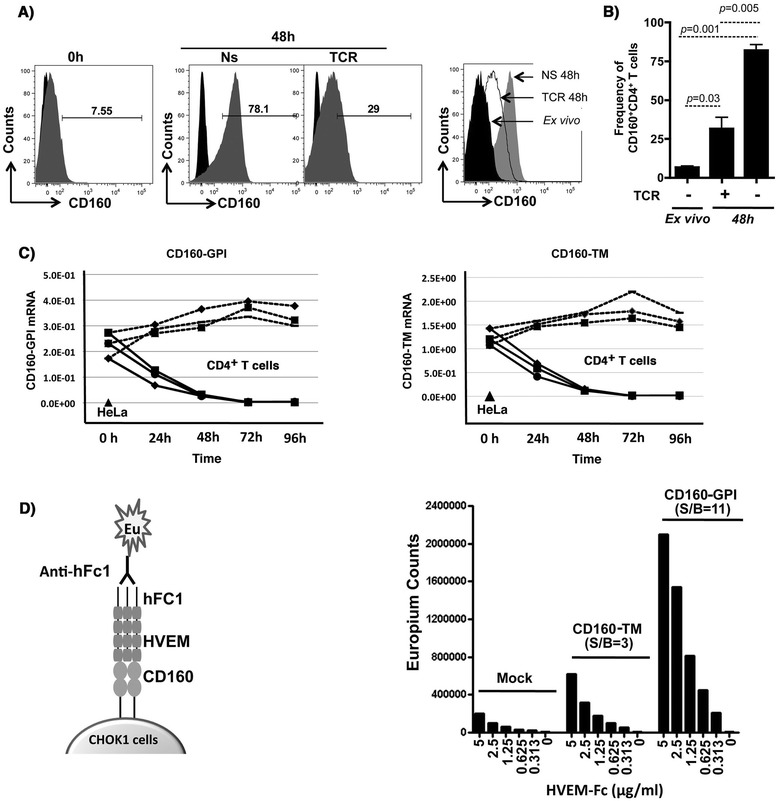
Figure 1.

Expression of CD160 isoforms in primary CD4 + T-cells and binding to HVEM. A) Left panel: Representative FACS analysis of CD160 on primary CD4+ T-cells isolated from total PBMCs of a healthy donor (ex vivo at baseline), gated on CD3+CD4+CD8− cells. Middle panels: CD160 surface expression following 48 h of resting (non-stimulated, NS) or TCR activation (plate-bound anti-CD3 and soluble anti-CD28). Right panel: overlapping histograms showing CD160 surface expression from TCR-stimulated CD4+ T-cells (dotted empty histogram) in comparison to 48 h rested CD4 (filled grey histogram) and freshly isolated CD4 cells (filled black histograms) all from the same individual donor. B) Frequency of CD160+CD4+ double positive population following 48 h of resting or TCR stimulation compared to freshly isolated (ex vivo) cells (n = 3). C) Kinetics of CD160-GPI and CD160-TM isoform expression at the mRNA level by quantitative RT-PCR in primary CD4+ T-cells (cells from n = 3 independent healthy donors) stimulated through TCR for 4 days. Values are relative to the house-keeping GAPDH gene transcripts (n = 3). HeLa cells were used as a negative control for CD160 TM transcription. D) Binding of HVEM to the two isoforms CD160. Left panel: Schematic representation for the TRF binding assay between CD160 (over-expressed by CHO-K1 cells) and the soluble ligand HVEM containing the human Fc1 (detection with anti-human Fc1). Right panel: Measuring the signal/background (S/B) for HVEM binding to both CD160-GPI and CD160-TM cells by the TRF assay under decreasing concentrations of HVEM-Fc.
