Figure 5.

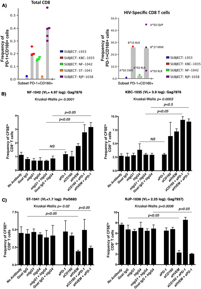
Enhanced CD8 + T-cell proliferation to antibody-mediated blockade of PD-1 in combination with either CD160 or HVEM antibodies. A) Histogram summarizing the phenotypic analysis showing the frequencies of CD160+PD-1+ double positive population on total CD8 (left panel: each dot represents an independent staining from the same subject) and HIV-specific (right panel) T-cells from the four recruited study subjects. L933 represents the HIV-uninfected donor used as a control. HIV pentamers from each subject is annotated above each bar (right panel). Gating was done on CD3+ lymphocytes followed by gating on either total CD8+ T-cells or pentamer HIV-specific CD8+ T-cells. Note that we were unable to fold the peptides identified in the ELISPOT assay with HLA-restricted multimers from cART-treated subject. B) CFSE lymphoproliferation assays on total PBMCs from the viremic subjects NF-1042 and KBC-1035 gated on CD3+CD4−CD8+ T-cells. PBMCs stimulated or not with Gag7876 (restricted by HLA-B*1501 for KBC-1035, and HLA-A*0301 for NF-1042) in the absence or presence of blocking antibodies (4 replicates for each condition). C) PBMCs from ST-1041 and RJP-1038 stimulated or not with Pol5683 (restricted by HLA-A*11, A*03, A*68) and Gag7937, restricted by HLA-B*2705, respectively (4 replicates for each condition). P values were determined using the nonparametric Kruskal-Wallis and Dunn’s post-test.
