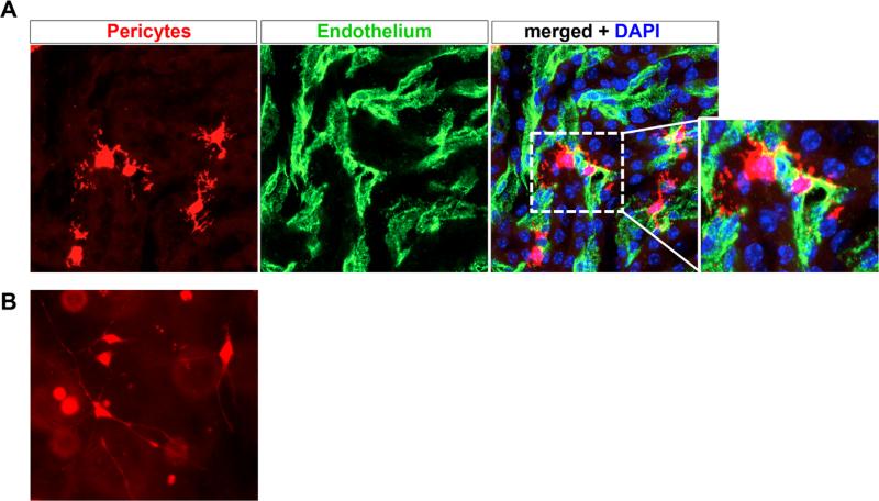
Figure 1.
Renal pericytes associate closely with vasculature. (A) Col1α1-CreERt2 mice were crossed against R26RtdTomato reporter mice, and pulsed with tamoxifen to induce recombination in medullary pericytes. A high-power view of tdTomato+ pericytes is shown, note the extensive branching processes emanating from the cell body. Endothelial cells were co-stained green using fluorescein isothiocyanate FITC-CD31 antibodies, and the merged image shows how tightly the pericytes associate with and wraparound peritubular capillaries. Genetically tagged renal pericytes (left panel) surrounding peritubular capillaries (middle panel, CD31). (B) Genetically labeled pericytes were dissociated from kidney by enzymatic dissociation and purified by fluorescence activated cell sorting. Subsequently, they were plated in a three-dimensional collagen gel, where long delicate processes extend from the cell body, similar to the in vivo situation. DAPI, 4',6-diamidino-2-phenylindole.

