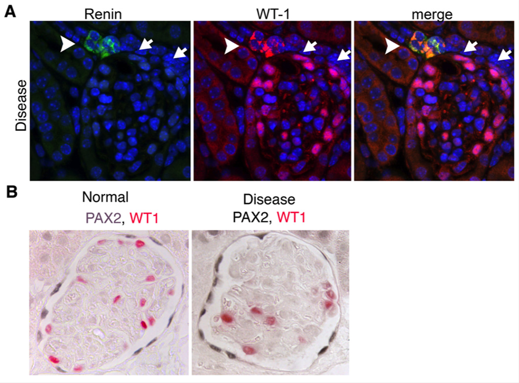
Figure 3. Renin producing vascular smooth muscle cells activate WT1 and PECs upregulate PAX2 in adult glomerular disease with podocyte loss.
(A) Split panel images of a glomerulus following administration of anti-podocyte antibodies showing de novo expression of WT1 in the renin producing cells of the JGA (arrowhead). In addition to expression of WT1 in podocytes attached to the tuft, WT1 can also be seen in some PECs (arrows). (B) Images of normal and diseased glomeruli following anti-podocyte antibody administration. Note increased expression intensity of PAX2 in PECs

