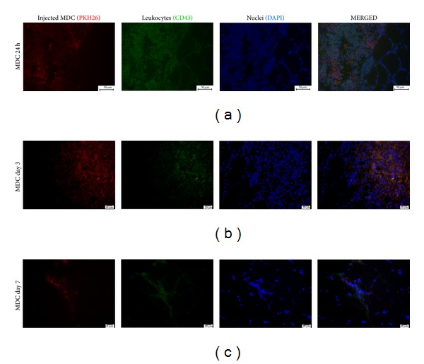
Figure 5.

Immunohistochemical staining in different time points. Leukocytes (CD43 antigen stained with Alexa 488 (green)) are infiltrating the cluster of transplanted MDC (PKH26 (red)). Cell nuclei stained with DAPI (blue) in different time points. Scale bars: 50 μm.
