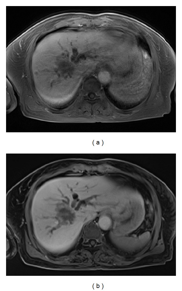
Figure 4.

MR images obtained in a 70-year-old woman with an intrahepatic mass-forming cholangiocarcinoma. The T1-weighted 3D conventional breath-hold GRE image (a) shows blurred tumor resolution and dilated intrahepatic bile ducts caused by moderate motion artifacts. The free-breathing T1-weighted 3D radial GRE image (b) shows clear definition of the liver tumor and renders increasing conspicuity of bile-duct invasion. No motion artifacts are present in this image.
