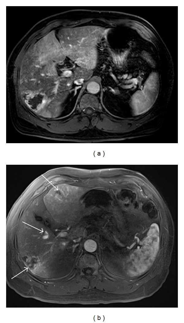
Figure 8.

MR images obtained in a 61-year-old man with multiple hemangiomas. (a) A hepatic arterial dominant phase image using conventional 3D GRE during a 20-second breath-hold shows respiratory motion artifacts. (b) A hepatic arterial dominant phase image using the CAIPIRINHA 3D GRE sequence during a 12-second breath-hold demonstrates good image quality without artifacts, high spatial resolution, and a well-timed late arterial image and, thus, resulting in detecting an increasing number of hemangiomas (arrows).
