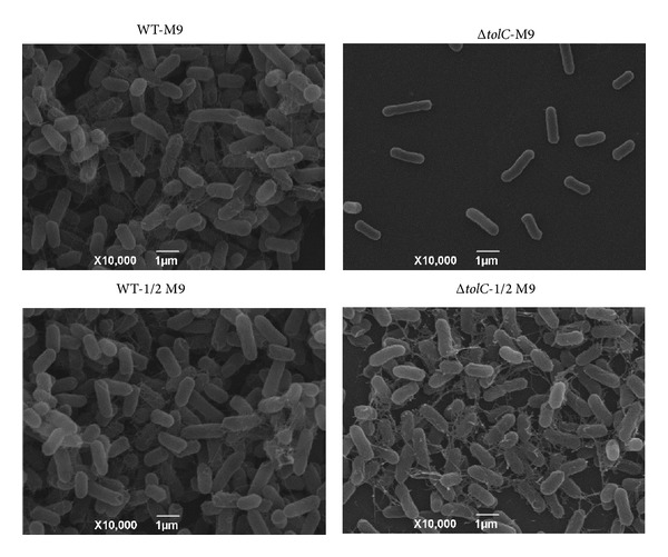
Figure 3.

Scanning electron microscopy (SEM) images of the WT strain (left) and ΔtolC strain (right). All strains were incubated on glass coverslips at 28°C for 5 days in M9 medium (upper) or 1/2 M9 medium (lower) and viewed under 10,000x magnification.
