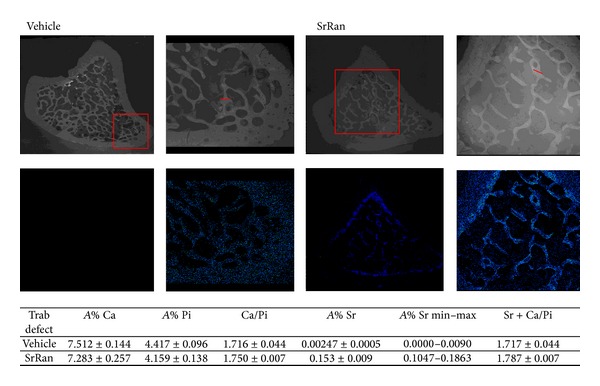
Figure 5.

X-Ray spectroscopy of proximal tibia cross-section of two representative specimens following 12 weeks of vehicle or SrRan administration. Squares show strontium distribution in bone tissue in greater detail. Strontium deposits in both newly formed cortical and trabecular bone in SrRan treated rats, including the defect area. In the table, the atomic percent averages of Sr, Ca, and Pi were determined in profiles along trabecular units at the edge of the defect and are depicted by straight lines in the figure.
