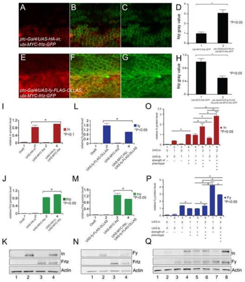
Figure 6.
The over expression of one PPE protein affects the accumulation of the others. A, B and C show a ptc-Gal4/UAS-HA-in; ubi-myc-frtz-GFP/+ wing immunostained for both In (red) and GFP (green-Frtz). This ubi-myc-frtz-GFP transgene gives variegated expression. D shows the quantitation of GFP (Frtz) staining inside (2) and outside (1) of the ptc domain. (Note the quantitation was done on the peripheral accumulation in cells that expressed high levels of Frtz). The increase in Frtz accumulation was significant (p<0.05). E, F and G shows a ptc-Gal4/UAS-fy-Flag-ollas; ubi-myc-frtz-GFP/+ wing immunostained for both Ollas (red-Fy) and GFP (green-Frtz). H shows the quantitation of GFP (Frtz) staining inside (2) and outside (1) of the ptc domain. The decrease in Frtz accumulation was significant (p<0.05). I, J and K show the results of Western blots of wing disc samples where the expression of UAS-HA-in and UAS-myc-frtz were driven either singly or together using ptc-Gal4. I and J show the quantitation and K the blot. M, N and L show the results of Western blots of wing disc samples where the expression of UAS-fy-Flag-Ollas and UAS-myc-frtz were driven either singly or together using ptc-Gal4. L and M show the quantitation and N the blot. O, P and Q show the results of Western blots of wing disc samples where the expression of UAS-HA-in and UAS-fy-GFP were driven either singly or together in varying doses using ptc-Gal4. O and P show the quantitation and Q the blot.

