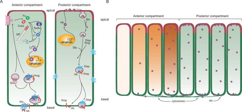
Figure 3. Model for Hh signaling and distribution in polarized Drosophila disc cells.

(A) Drawing depicts a model of production and signal transduction in Hh-producing Posterior compartment and Hh-responding Anterior compartment cells. In Hh-producing cells, Hh (purple) that has lipid modifications (red and green circles) is packaged in vesicles that invaginate in a Shibire-dependent (Shi) process from the apical plasma membrane. These vesicles incorporate Ihog (orange) and Dispatched (Disp, blue) and deliver vesicular Hh to the basal plasma membrane. Hh is taken up basally by Hh-responding cells, generating vesicles that contain Ptc and Ihog. Smo redistributes from intracellular vesicles to the plasma membrane and interacts with components of Hh signal transduction (Fused (Fu), microtube (MT)-associated Costal-2 (Cos2) and Su(fu)) to both inhibit production of the repressor form of Ci (CiR) and induce production of the activator form of Ci (CiAct). (B) Drawing depicts an epithelium with Hh-producing (right, green) and Hh-responding (left, orange) cells, and Hh transport (left to right) from Posterior compartment Hh-producing cells by basal cytonemes to Anterior compartment Hh-responding cells. Both the concentration gradient of Hh (purple spheres) and the concentration-dependent response (orange) in the Anterior cells are represented.
