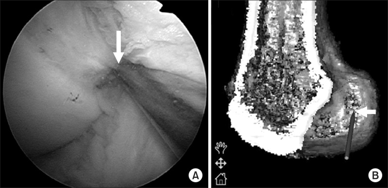
Fig. 3.
Arthroscopic and navigation views of the femoral guide. (A) Arthroscopic view of the lateral wall of the femoral intercondylar notch can be obtained through the anteromedial (AM) portal with the knee in 90° flexion. The tip of the femoral guide is placed within the anatomical femoral footprint through a far AM portal. (B) Navigation view of the lateral wall and roof of the femoral intercondylar notch on the three-dimensional reconstructed image. The white arrow shows the tip of the femoral guide.

