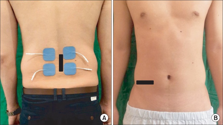
Fig. 1.
(A) Sites of stimulation and recording: lumbar paraspinal region and abdominal region shown in blue; 4 hydrogel surface electrodes (5 cm×5 cm) placed bilaterally at the level of the L4 and L5 spinous processes shown in black. (B) Site of the ultrasound transducer: the abdominal ultrasound transducer position was adjusted to ensure that the medial edge of transversus abdominis muscle was approximately 2 cm from the medial edge of the ultrasound image when the participant was relaxed.

