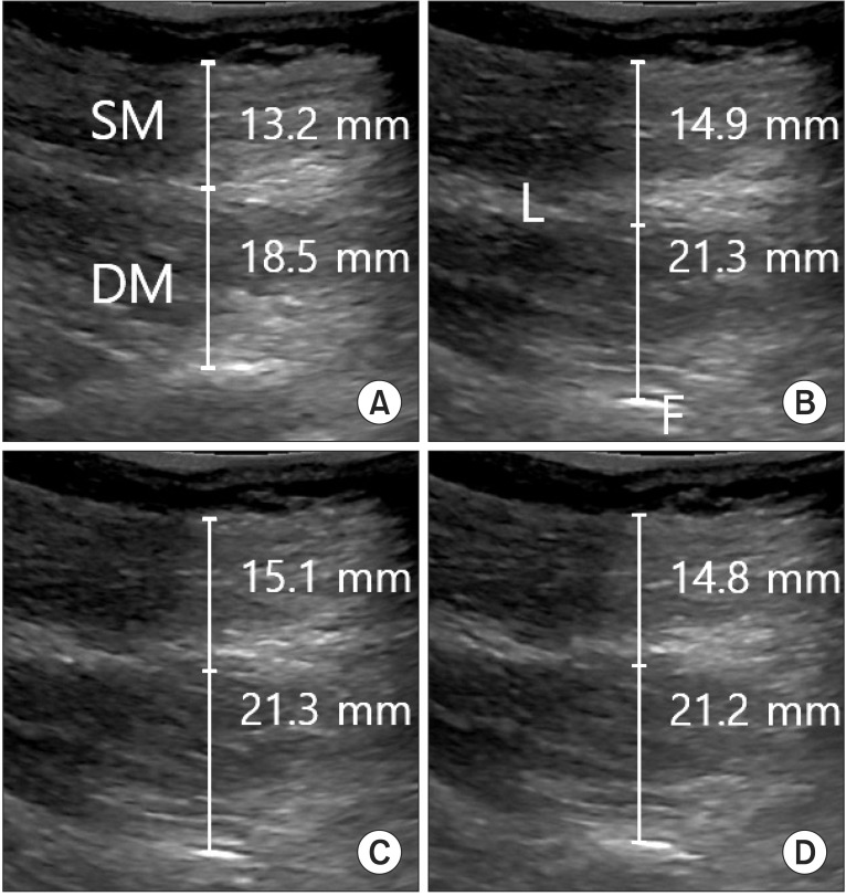
Fig. 3.
Comparison of the thicknesses of superficial and deep lumbar multifidus (LM) muscles during NMES. At rest (A), NMES at 20 Hz (B), NMES at 50 Hz (C), NMES at 80 Hz (D). LM thickness was measured between the posteriormost portion of the L4-5 facet joint ('F' in B) and the fascial plane between the muscle and subcutaneous tissue. SM and LM were separated along the edge of the hypoechoic region ('L' in B), which represents the locations of fascial separations between muscles. NMES, neuromuscular electrical stimulation; SM, superficial multifidus; DM, deep multifidus.

