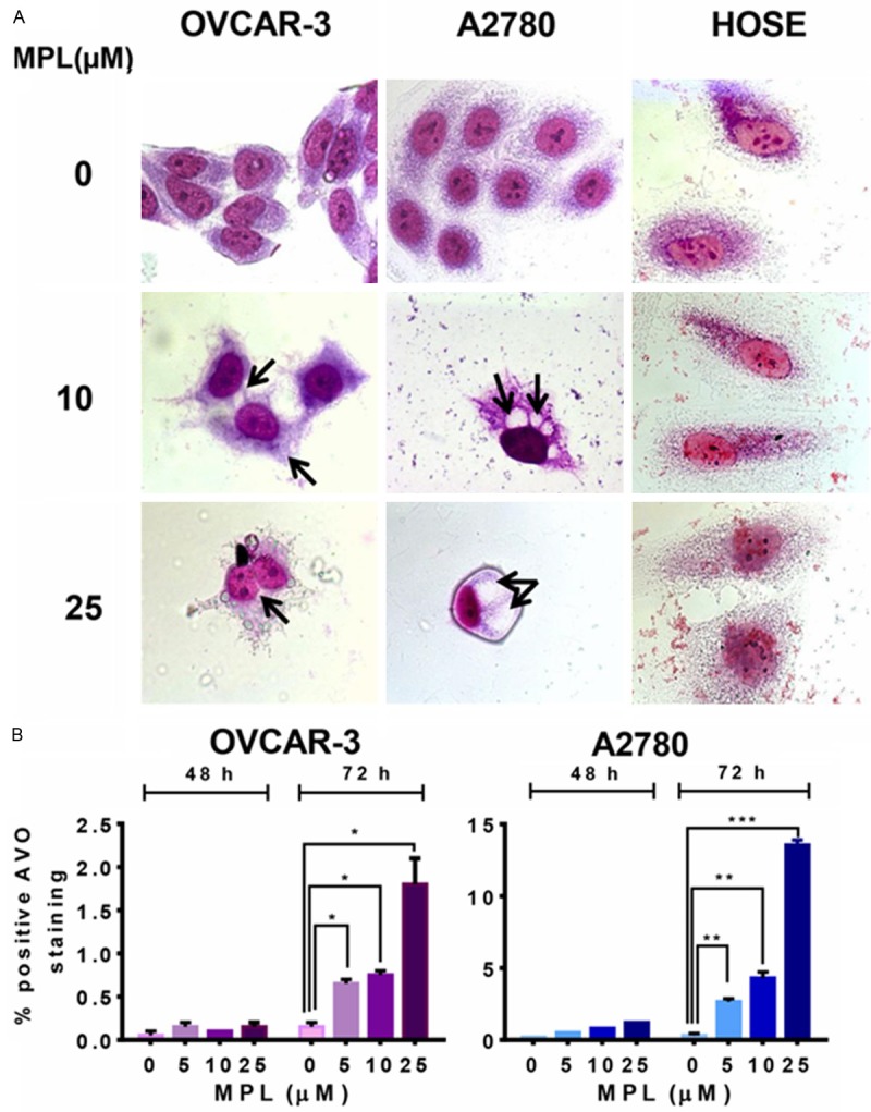
Figure 2.

MPL induces accumulation of acidic vacuoles. A. Human ovarian cancer cell lines (OVCAR-3 and A2780) and Normal human ovarian surface epithelial (HOSE) were grown in six-well tissue culture plates under standard cell culture conditions in the presence of MPL (0, 10, 25 μM) for 72 h. Cells were then stained with giemsa, washed and photographed under Leica DM IRB light microscopy (magnification 100x) with oil immersion lenses. A representative from two independent experiments has been shown. B. The percentage of bright red fluorescence (FL3)-positive cells was evaluated by AO staining and FACS analysis in OVCAR-3 and A2780 cells untreated or treated with MPL (0, 5, 10, 25 μM) at different time points (48 and 72 h). Results are the mean ± SEM of duplicate samples from one experiment and repeated three with similar results experiments.
