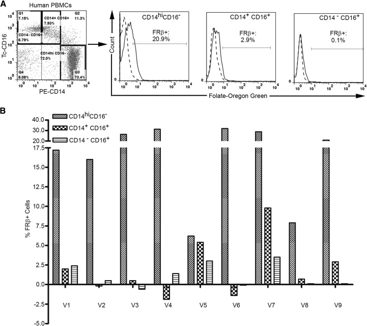
Figure 4. Binding of a folate-conjugated fluorescent probe to human peripheral blood monocytes.
(A) Human PBMCs were incubated with 250 nM FOG for 1 h at 37°C and stained with antibodies to CD14 and CD16, followed by washing extensively with PBS to remove unbound labels. Uptake of FOG by three major subsets of human monocytes (CD14highCD16−, CD14+CD16+, CD14−CD16+) was then evaluated in the absence (solid lines) and presence (dashed lines) of 1000× molar excess of free folic acid to block all empty FRs. (B) Summary of the expression of functional FR-β on the above subsets of human peripheral blood monocytes from nine volunteers (V1–V9).

